
Here are the most common types treated in specialized wound care settings
Ulcerations (or ulcers) are open sores that result from the breakdown of skin or mucous membrane tissue. In wound care, especially at a clinic like Oregon Coast Wound Center, we focus primarily on chronic lower extremity ulcers — those that don’t heal within weeks and often stem from underlying conditions like diabetes, poor circulation, or pressure.
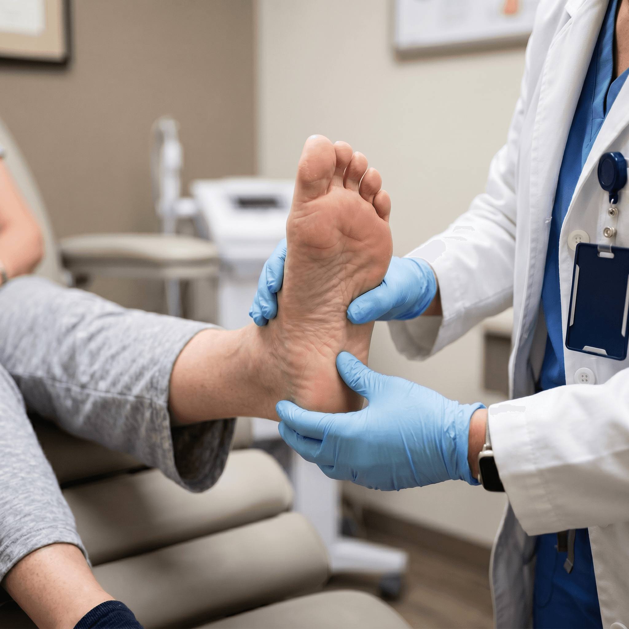
1. Diabetic Foot Ulcers (Neuropathic Ulcers)
Causes:
Primarily peripheral neuropathy (nerve damage from high blood sugar) combined with poor circulation and repetitive trauma/pressure. Patients often can’t feel pain, so small injuries worsen unnoticed.12
Bottom of the foot (pressure points like heels, ball of foot, or toes), but can occur anywhere on the foot.
Appearance:
ften painless, with a punched-out or crater-like look. May have callused edges, moderate drainage, and surrounding redness if infected. Granulation tissue may be present but healing is slow.
Risks:
High chance of infection, osteomyelitis (bone infection), and amputation if not managed aggressively.
Treatment Focus:
Offloading (reducing pressure with casts, boots, or custom footwear), debridement, infection control, blood sugar management, and advanced therapies like bioengineered grafts or NPWT.
2. Venous Ulcers (Venous Stasis Ulcers)
Causes:
Chronic venous insufficiency — damaged vein valves cause blood to pool in the legs, leading to high pressure, swelling, and tissue damage.
Typical Location:
Lower leg, often around the inner ankle (medial malleolus).
Appearance:
Shallow, irregular borders with a “weeping” or moist base. Surrounding skin may be discolored (brownish hemosiderin staining), itchy, or have varicose veins/edema. Usually less painful than arterial ulcers but can ache with dependency.
Risks:
Recurrence is common (up to 70% without proper compression); infection and cellulitis.
Treatment Focus:
Compression therapy (the cornerstone), leg elevation, wound cleaning, advanced dressings, and addressing underlying vein issues. NPWT or skin substitutes help in stubborn cases.
3. Arterial Ulcers (Ischemic Ulcers)
Causes:
Poor arterial blood flow (peripheral artery disease/PAD), often from atherosclerosis, smoking, diabetes, or high blood pressure. Tissues don’t get enough oxygen and nutrients
Typical Location:
Toes, feet, or areas exposed to trauma (shins, heels). Often on the outer ankle or pressure points.
Appearance:
Deep, “punched-out” with well-defined edges. Pale or necrotic (black) base, minimal drainage, and little to no granulation tissue. Very painful, especially at night or when legs are elevated. Surrounding skin is often shiny, hairless, and cool.
Risks:
Rapid worsening, gangrene, and high amputation risk without revascularization.rrence is common (up to 70% without proper compression); infection and cellulitis.
Treatment Focus:
Vascular assessment and possible referral for angioplasty/bypass, pain management, careful debridement (only after improving blood flow), and protective dressings. Avoid compression.
4. Pressure Ulcers (Pressure Injuries / Bedsores / Decubitus Ulcers)
Causes:
Prolonged pressure on skin over bony areas, often combined with shear, friction, moisture, and poor nutrition. Common in immobile patients, elderly, or those with limited mobility.
Typical Location:
Sacrum/tailbone, heels, hips, elbows, or any bony prominence.
Appearance:
Ranges from non-blanchable redness (Stage 1) to full-thickness tissue loss exposing muscle or bone (Stage 4). Can include deep tissue injury. May have slough or eschar.
Risks:
Infection, sepsis, prolonged hospitalization.
Treatment Focus:
Pressure offloading (special mattresses, repositioning), nutrition support, debridement, moisture management, and advanced therapies. Prevention is key.
Mixed or Other Ulcers
Many patients have mixed etiology ulcers (e.g., diabetic + arterial, or venous + arterial components). Accurate diagnosis through vascular testing (ABI, Doppler), imaging, and clinical exam is essential.
Less common types include:
Traumatic ulcers (from injury)
Malignant ulcers (cancer-related)
Infectious ulcers (e.g., from osteomyelitis)
Why Differentiation Matters
Each type requires a different treatment approach. Using compression on an arterial ulcer can worsen it, while ignoring offloading in a diabetic ulcer leads to failure. At Oregon Coast Wound Center, we start with a comprehensive assessment (vascular checks, wound measurement, labs) to create a personalized plan.
Prevention Tips (relevant for coastal patients):
Daily foot checks (especially if diabetic)
Blood sugar and vascular health management
Proper footwear and compression when indicated
Mobility and nutrition support
If you or a loved one has a non-healing wound, early evaluation dramatically improves outcomes and reduces complications.
